What Can I Expect from a Retina Specialist?
A retina specialist is a medical doctor who has specialized in ophthalmology and sub-specialized in diseases and surgery of the vitreous body of the eye and the retina.
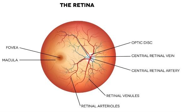
A retinal specialist focuses their work on the retina, which is the delicate nerve tissue that lines the back inside wall of the eye, the vasculature which supports the retina, and the vitreous which is the jelly material that fills the central cavity of the eye.
When you visit a retina specialist, you can expect to have a retinal examination — sometimes called ophthalmoscopy or funduscopy — which allows your specialist to evaluate the back of your eye, including the retina, the optic disk and the underlying layer of blood vessels that nourish the retina (choroid).
During a retinal exam and consultation in Phoenix, our doctors will perform a series of diagnostic procedures in order to evaluate the retina for any sign of disease or abnormality.
These tests may include:
- Fundus Photography: Fundus photography uses specialized film and digital cameras to document abnormalities in the retina. It is important in following the progress of certain retinal diseases and to monitor treatment.
- Fluorescein Angiography: Fluorescein angiography is a test used to examine blood vessels in the retina. This procedure involves the injection of a vegetable-based fluorescein dye into the blood stream. As the blood circulates through the retina, a series of rapid, sequential photographs are taken of the eye. This commonly performed test provides useful anatomic and functional information about the retina and is one of the most important tools in the diagnosis and treatment of retinal disorders.
- OCT: Optical Coherence Tomography (OCT) is a non-invasive imaging technique relying on low coherence interferometry to generate cross-sectional imagery of ocular tissues. Cross-sectional visualization is an extremely powerful tool in the identification and assessment of retina abnormalities.
- Visual Field: Visual field testing is a sophisticated, automated computerized vision test that measures both central and peripheral vision/visual function. It is essential in monitoring glaucoma and it is a useful ancillary test to diagnose, document or treat neurologic or retinal disease.
- B-scan Ultrasound: B-scan ultrasound is a method for viewing the structures at the back of the eye through the use of high frequency sound waves. This technology provides a topographical view of the eye with information that is not otherwise possible.
Our doctors will take the time to discuss the results of your exam, as well as any potential risks of retinal disease with you. Patient education and understanding is a top priority of Associated Retina Consultants, as it often helps patients achieve the most successful outcome.
To schedule a consultation with a retina specialist in Phoenix, contact Associated Retina Consultants at 602-242-4928 or website.